|
Ligamentous injury to the knee usually results from an indirect force such as a twist or varus or valgus force on the knee. Check for distal pulses and neurological injury such as a drop foot, signifying a possible injury to the peroneal nerve. The presence of a heamarthrosis may mean that the injury did not significantly tear the capsule. The stability of the knee ligaments are tested by stressing them. X rays are taken to show any ligamentous avulsions or more serious fractures such as a Condylar or plateau fracture.
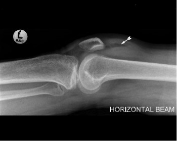
A horizontal shoot through film may show a lipohaemarthrosis - a radiolucent area in the suprapatellar sac representing fat floating on the blood in the knee. If examination is difficult because of pain, aspiration of the heamarthrosis together with injection of local anesthetic may permit adequate evaluation of the ligaments. It is advisable to aspirate a heamarthrosis if it is causing tension in the knee, small intraarticular bleeds may be ignored. Examine the aspirated fluid for fat droplets. These may signify the presence of an intraarticular fracture. Knee DislocationsA dislocated knee is obvious while a severely injured knee, with multidirectional instability may have been dislocated, and has since spontaneously reduced. Regard all severe knee injuries as dislocations. Check and note the presence or absence of foot pulses. Up to 30% of knee dislocations have vascular compromise. Check for peroneal nerve injury by asking the patient to dorsiflex the foot.
Management:Reduce the knee immediately.It usually reduces easily with only sedation being necessary. If difficulty is encountered open reduction in theatre will be needed. Obtain an arteriogram to check the vascular status even if pulses are intact. If there is vascular compromise an emergency vascular repair will be needed. A grossly unstable knee can be temporarily stabilised with an exfix over the knee. Conservative treatment consists of an above knee plaster cast for six weeks. Because of the scarcity knee dislocations and the variety of ligaments torn, it is still unproven whether surgical repair of ligament is superior to plaster immobilisation. The consensus is that surgical treatment is better. Ligamentous repair is usually delayed for about a week. All major ligaments are sutured. The anterior and or posterior cruciate ligaments are usually damaged in a dislocation. In substance tears need replacement by either a bone patellar bone or hamstring substitutes. "Isolated" Ligamentous rupturesMore minor knee ligament disruptions are common in contact sports.
Milder knee injuries can be treated conservatively with a Robert Jones bandage for one to 6 weeks, depending on the severity of the injury. Once pain has subsided physiotherapy to restore range of motion and build up the quadriceps muscle is required. If x rays show avulsion fractures these may favor surgical repair as avulsed ligaments can easier be repaired than a mid substance tear. Surgical repair of ligaments is usually done within 14 days of injury. If the cruciates require repair by means of a substitute (bone patellar bone, or hamstring) it may be wise to allow other ligaments to recover, and to first rehabilitate the knee with physiotherapy. A mobile knee is vital to the success of this type of surgery. The surgeon can then repair the cruciates on a fully rehabilitated knee at 6 weeks or more post injury. Meniscal tearsSuspect a tear of the meniscus if the patient complains of "locking" or inability to extend the knee. There is usually a history of a previous injury. The classical injury is: O' Donahague's triad:
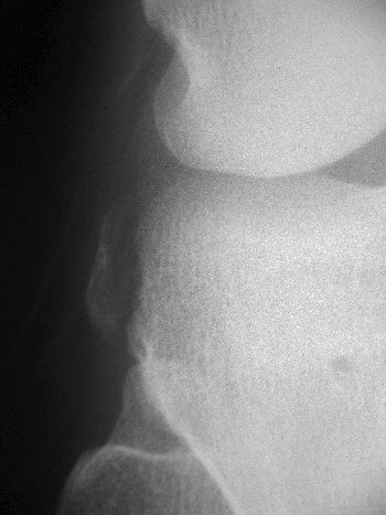
More commonly it is the lateral collateral of the knee that gives way first with an anterior cruciate ligament rupture, with, or without a meniscal tear. A sure sign of an anterior cruciate ligament rupture is the Segond avulsion. A avulsion fracture on the lateral side of the tibial plateau.
Treatment of meniscal tears. The meniscus seldom heals on its own. Arthroscopic partial menescetomy will be required.
|