|
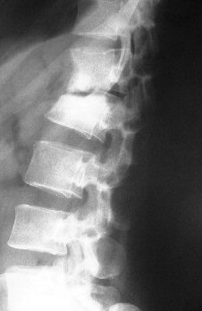
| Note the loss of disc height. There is also soft tissue swelling anteriorly to the involved vertebrae. |
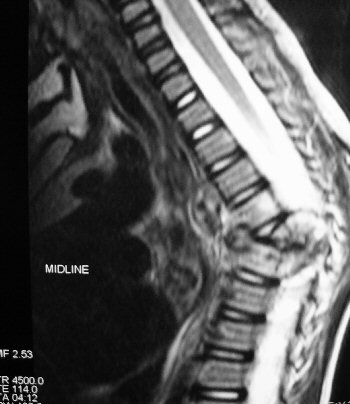 |
The magnetic resonance shows compression of the spinal
cord |
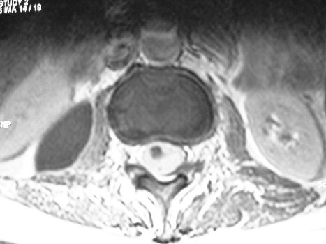 |
MRI: A "cold abscess" of TB has increased the signal
in the left psoas |