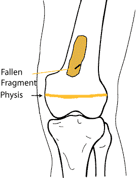
Simple bone Cyst
Eccentric, non expansile. One compartment. May see "fallen fragment " sign. Starts at physis and migrates away with growth. Has a long axis aligned to the bone shaft. Never penetrates cortex to extend into soft tissues.
|
Simple bone Cyst Eccentric, non expansile. One compartment. May see "fallen fragment " sign. Starts at physis and migrates away with growth. Has a long axis aligned to the bone shaft. Never penetrates cortex to extend into soft tissues. |
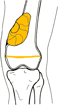 |
Aneurysmal Bone Cyst Usually metaphyseal, may extend into epiphysis Blown out appearance with thinned outer cortex. Multi compartmental. |
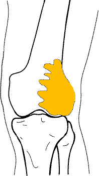 |
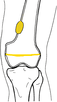
Giant Cell Tumour Seen in adults. Extends to the subchondral bone of the joint margin. Crosses from metaphysis into epiphyseal region. May be expansile. Outer border may be thinned or absent (may break into soft tissue). |
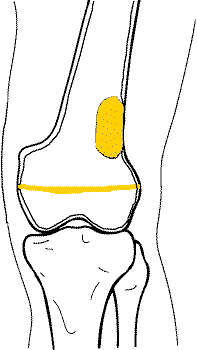 |
Non Ossifying Fibroma / Fibrous Cortical Defect Eccentric and attached to cortex. Commonly in metaphysis, but may be seen in shaft. Well defined smooth or scalloped margins. |
 |
Chondromyxiod Fibroma Eccentrically situated lytic lesion with well defined margins in the metaphysis of the lower extremity. The lesion usually has a sclerotic margin of bone and a lobulated contour. Ridges and grooves that appear in the margins secondary to scalloping falsely appear to be trabeculae |
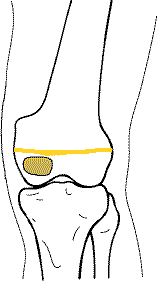 |
Chondroblastoma Characteristically involves the epiphysis.Well demarcated oval or round radiolucency. Has a thin sclerotic bony margin. Fine calcifications, either punctate or in rings, may be visible. |