Play hip dislocation video slideshow
Posterior Hip Dislocations
Posterior hip dislocations are the most common. The usual cause is a motor vehicle accident with the passenger's knee hitting the dashboard and forcing the femoral head out of the acetabulum posteriorly. The limb is shortened, and the hip flexed, the foot is in internal rotation.
Check for other fractures especially femur neck or shaft fractures and tibial fractures. Remember that associated fractures of the ipsilateral femoral shaft are not uncommon.
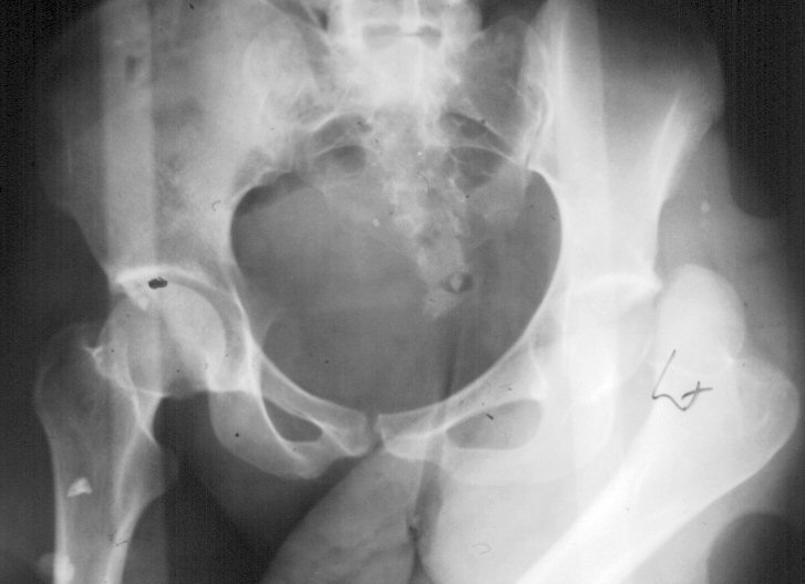 |
X-ray signs posterior dislocation
|
Obtain a pelvic x ray when managing any femur or tibia fracture, especially in the unconscious victim.
Associated femur head and acetabular rim fractures of ten complicate these injuries. Sciatic nerve damage is common - check that there is good dorsiflexion of the foot.
Reduction - posterior dislocation
Reduction is an emergency - do not delay this. Closed reduction and should
be performed at the original hospital if it is any significant distance
from the regional institution as delay significantly increases the incidence
of vascular necrosis. 
Use general anaesthetic with muscle relaxants. An assistant steadies the pelvis while the surgeon applies longitudinal traction to get the femoral head under the acetabulum. The hip is now flexed and pulled upwards. The internal rotation is corrected at this stage and a click should be felt as the hip reduces. Once reduced check the hip for stability. It should have a full range of motion and be in neutral rotation. Obtain a postoperative x ray to confirm the hip is concentrically reduced. Widening of the joint space may mean that there are bony fragments in the joint cavity (later arthrotomy and repair may be needed) or that the acetabulum is deficient due to a fracture.
The patient is placed in skin traction or a Denham pin may be inserted into the proximal tibia for 3 to 6 weeks traction.Any femur head and acetabular fractures are operated on electively, once computer tomogrammes are done to define the lesion.
Complications Posterior hip dislocation |
|
Anterior Hip Dislocations
With an anterior dislocation the lower limb is lengthened, the hip abducted and the foot is in external rotation.
 As the femur
head is either anterior in the groin or in the obturator fossa it can
obstruct the femoral vein causing thrombosis and possible pulmonary embolism.
As the femur
head is either anterior in the groin or in the obturator fossa it can
obstruct the femoral vein causing thrombosis and possible pulmonary embolism.
X ray signs of an anterior hip dislocation are the lesser trochanter being more visible (due to external rotation. The hip is abducted and the femur head is usually inferior to the acetabulum. Shenton's line is also broken.
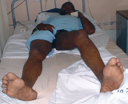 |
| Anterior dislocation. Hip is in abduction. |
Reduction - anterior hip dislocation
You will need at least one assistant and an anesthetist to reduce an anterior hip dislocation. Under general anaesthetic the assistant disimpacts the femur from the obturator fossa by applying a lateral force to the hip.
